Knee Dislocations: Recognition, Emergency Care, and Recovery (Canada)
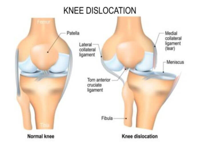
A knee dislocation occurs when the end of the femur (thighbone) is no longer aligned with the tibia (shinbone). This is a serious medical emergency that requires immediate assessment in a hospital setting, as it can damage major blood vessels and nerves in the leg.
In many cases, a knee dislocation happens when the lower leg is forced beyond the normal range of the knee joint. The shinbone may be pushed forward, backward, or to either side of the thighbone. In Canada, most knee dislocations are linked to high-energy trauma, such as motor vehicle collisions, workplace incidents, or sports injuries. However, in some individuals, even a seemingly minor event—such as stepping into a hole while twisting the knee—can result in a dislocation.
What Are the Indications of a Knee Dislocation?
In most cases, a knee dislocation is obvious. Common signs and symptoms include:
-
The knee appears visibly out of place or deformed
-
Severe pain and swelling
-
Inability or great difficulty walking or bearing weight
-
Instability of the joint
In some situations, the knee may slip back into place on its own before medical care is reached. Even if this happens, the injury is still very serious.
⚠️ Urgent warning signs include:
-
Numbness in the lower leg or foot
-
Pale or cool skin below the knee
-
Weak or absent pulse in the foot
These symptoms may indicate damage to an artery or nerve, which can threaten the viability of the limb if not treated promptly.
Diagnosing Knee Dislocations
If a knee dislocation is suspected, the individual should be taken to the nearest emergency department immediately.
A physician can often identify a dislocated knee through physical examination, but imaging is essential. Diagnostic steps typically include:
-
X-rays taken from multiple angles to confirm the dislocation and identify fractures
-
Repeated checks of circulation, including pulses in the lower leg
-
Assessment for nerve damage, by asking the individual to:
-
Move the foot up and down
-
Turn the foot inward and outward
-
Report any numbness or tingling
-
Even if the joint has already returned to its normal position, these assessments are critical to rule out hidden injuries.
Management and Treatment
Treatment of a knee dislocation is performed in a hospital setting.
-
The joint is carefully repositioned (reduced) back into place
-
The individual is given pain medication and a sedative, but usually remains conscious
-
After reduction, the knee is immobilized with a splint
If arteries are damaged, surgical repair is performed urgently to restore blood flow. Damage to ligaments, nerves, or other structures may also require surgery.
In cases where the knee remains highly unstable, an external fixator may be applied. This device consists of a frame and rods attached to the outside of the leg with metal pins inserted into the bone, helping stabilize the joint while healing begins.
Disclaimer:
Important – Educational Use Only (Canada):
This information is provided for general learning and first aid awareness. Knee dislocations are medical emergencies that can involve life-threatening vascular and nerve injuries. Do not attempt to realign a dislocated knee outside of a hospital setting. Immediate emergency medical care is required. Proper recognition, immobilization, and emergency response skills are taught in certified Canadian first aid and CPR courses.

